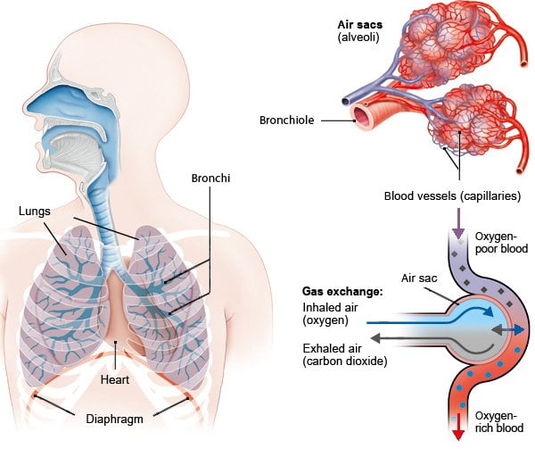
Our lungs are one of the largest vital organs in our bodies. The oxygen you breathe in enters your lungs and then enters your blood. It is then carried to all of your body’s cells via your bloodstream. The lungs are located in the chest region, where they are protected by the rib cage’s ribs. Their structure is comparable to that of an inverted tree: The windpipe divides into two airways known as bronchi, both of which connect to the lungs. Within the lungs, the airways continue to branch into narrower airways until they reach the air sacs.

How is the pulmonary circulation defined?
When you inhale (breathe in), oxygen-rich air enters your windpipe, travels through the bronchi, and finally reaches the air sacs. The alveoli, or air sacs, are responsible for gas exchange. They resemble grapes at the bronchial branch tips. A healthy lung contains approximately 300 million air sacs. Each air sac is encircled by a fine network of blood vessels (capillaries).
The oxygen in the inhaled air passes through the air sacs’ thin lining and into the blood vessels. This is referred to as diffusion. The oxygen in the blood is then carried by the bloodstream throughout the body, reaching every cell. Carbon dioxide is expelled from the bloodstream when oxygen is absorbed. CO2 is a byproduct of cellular metabolism. When you exhale, you expel it (exhale). This gas is transported in the opposite direction of oxygen: from the bloodstream through the lining of the air sacs to the lungs and out into the open.
What occurs when you inhale?
Your chest and lungs expand as you inhale. When you exhale, your lungs contract again. Both of these movements are facilitated by the diaphragm and the muscles running between the ribs (intercostal muscles). We breathe automatically.
Adults breathe 14 to 16 times per minute when at rest. During a normal breath, approximately half a liter of air is inhaled. When you become more active, your breathing becomes faster and deeper in order to oxygenate your blood more effectively.
A person’s overall fitness level is highly dependent on the efficiency of their lungs and heart. Numerous breathing tests can be used to assess your lung function.
Lung structure
The windpipe (trachea) is approximately ten centimeters long in adults and divides into two main bronchi known as the right and left bronchi. These principal bronchi then branch off into smaller secondary bronchi (lobar bronchi) – three in the right lung and two in the left lung. The left lung has less space because it shares space with the heart.
After that, the secondary bronchi branch into a variety of tertiary bronchi (segmental bronchi). The right lung is divided into ten segments referred to as bronchopulmonary segments. Nine of these segments comprise the left lung. Each segment is supplied with its own tertiary bronchus and pulmonary (lung) artery branch. This means that if necessary, for example, due to a serious lung disease or injury, individual segments can be removed.
Mucus-producing cells and millions of minute hair-like projections called cilia line the windpipe and bronchi. If you inhale harmful substances such as dust or other particles, the mucus and cilia prevent them from remaining in your lungs: Foreign matter becomes trapped in the mucus, and the cilia constantly move back and forth, transporting the mucus from the lungs to the throat, where it is swallowed or coughed out. A cough reflex is triggered when larger foreign objects enter the windpipe.

